


Image credit: Greg Moss / Wellcome Sanger Institute
We take an inside look into what laser capture microdissection is and how it is enabling scientists at the Wellcome Sanger Institute to precisely isolate cells and reveal detailed insights into biology and disease.
Imagine you are at a village fete. There is a stall with a steady-hand game: you guide a metal loop along a twisting wire, trying not to touch it and set off the buzzer, all to reach the end and win a prize. Now imagine playing that same game a million times smaller, where the margin for error is just a few micrometres. This is the daily challenge for scientists working with laser capture microdissection (LCM). Researchers must precisely guide a laser to cut around specific groups of cells, isolating them from the surrounding complex tissue, while leaving everything else untouched.
In this blog, we delve into what LCM actually is, why it is used and what information it enables our scientists to unlock in cancer biology and beyond.
What is laser capture microdissection?
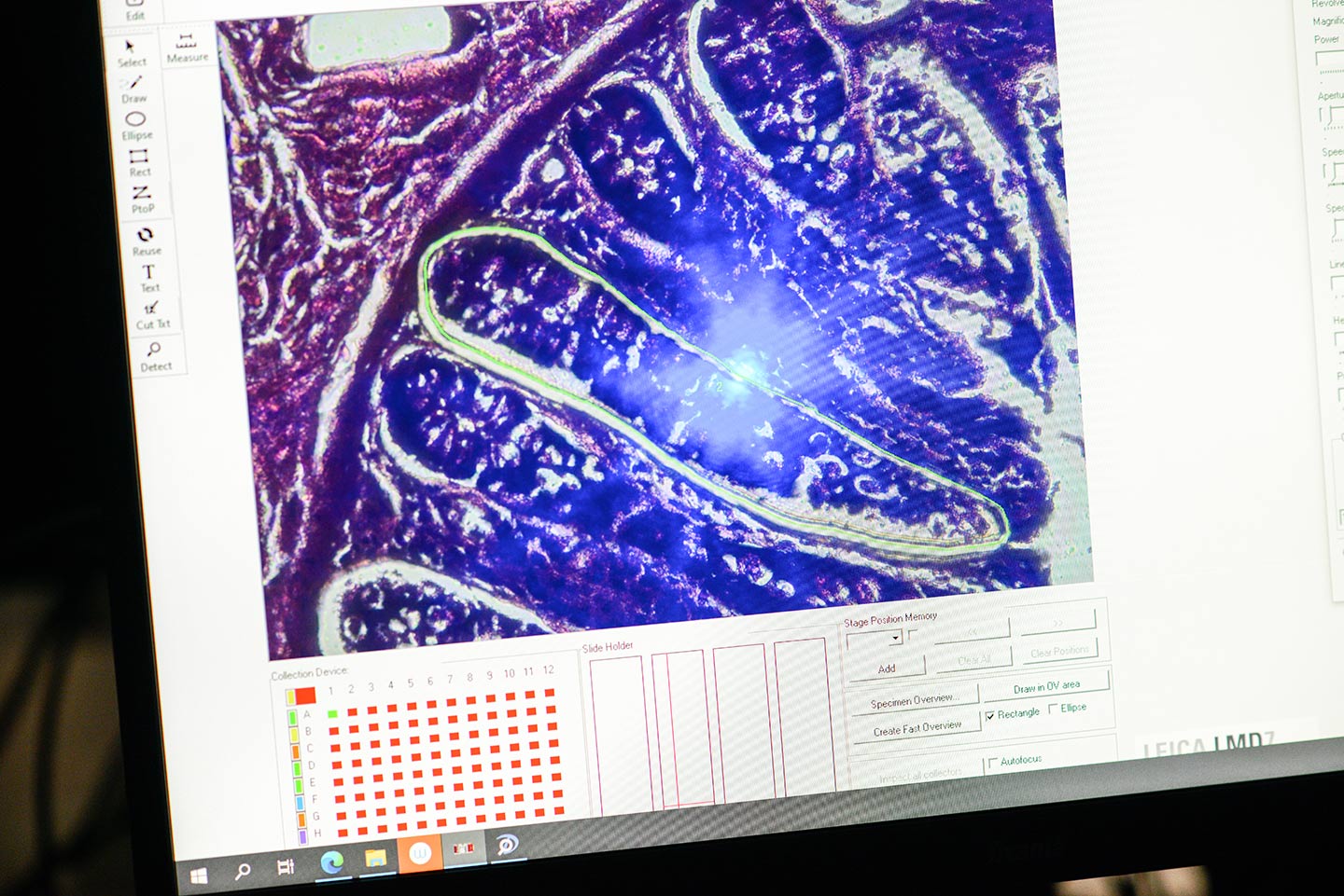
Laser capture microdissection (LCM) is a technique used to isolate specific cells or tissue regions from samples under a microscope. It utilises a near ultraviolet laser to cut around the region of interest. A pressure wave is then used to displace the tissue or cells into a collection tube. How this force is applied is critical: if focussed directly within the cut region, the tissue can move unpredictably, reducing both accuracy and recovery. Targeting the wave slightly above the tissue, however, pushes the sample downward more cleanly, improving collection reliability. This creates a two-step process: first, the region of interest is cut; second, the pressure wave is applied to collect it. Doing these steps separately gives you more control and helps you collect the material more reliably.

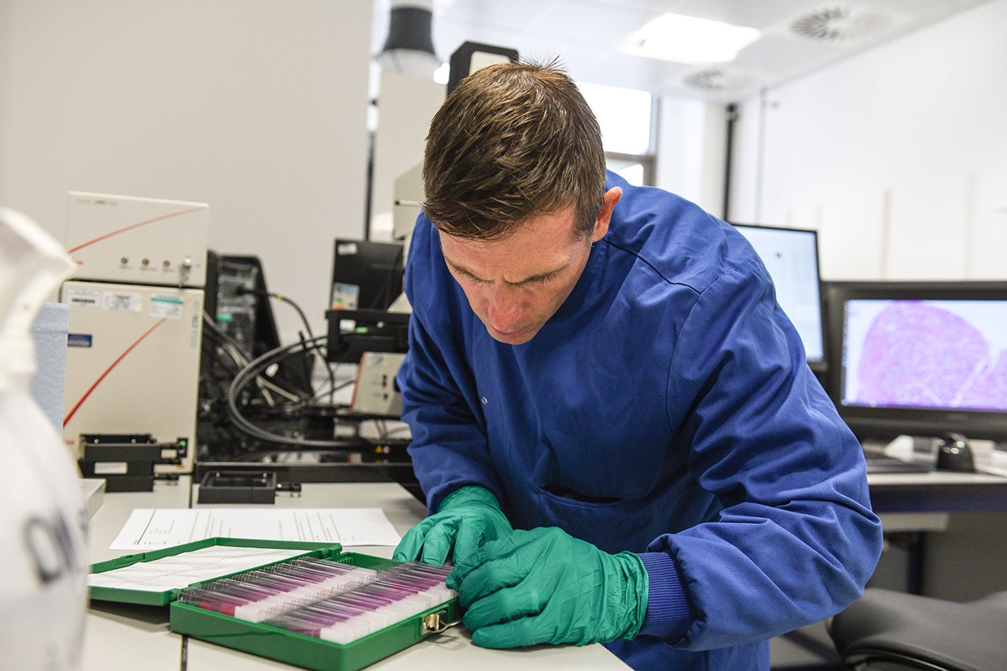
Paul Scott, Technical Specialist in the Somatic Genomics programme, is an expert in LCM with decades of hands-on experience, from early systems to today’s advanced platforms. He runs the LCM facility at Sanger, supporting a wide range of projects and training researchers to use the technology effectively. His expertise goes far beyond operating the equipment – LCM requires a deep understanding of the physics, biology and technique involved, where even small adjustments can make a big difference to results.
“Laser microdissection looks pretty straightforward, draw around the target, cut the tissue, then drop the isolate into your collection vessel. In reality, it is a highly sensitive technique; tissue type and condition, focus, the interaction between membrane, tissue and slide, and environmental factors such as humidity and static all have an impact. Getting LCM to work properly takes practice, understanding laser adjustment and positioning, correct application of pressure waves, and anticipation of sample behaviour make a huge difference.”
Paul Scott,
Technical Specialist, Wellcome Sanger Institute
History of LCM systems
Laser microdissection as a concept was first demonstrated in 1976 by German pathologists, who showed that laser energy could be used to target and manipulate tissue at a microscopic scale.1 However, it was not until the mid-1990s that LCM became a practical tool for isolating specific cells for molecular analysis.2 Researchers at the National Institutes of Health (NIH) developed modern LCM techniques around 1996, focussing on precise excision of cells while preserving their integrity for downstream studies.2
The first commercial LCM systems were then introduced by Arcturus in the late 1990s. These early systems featured a laser isolation mechanism and a collection method designed to capture cells with minimal contamination, though success rates were initially modest due to technical limitations and environmental sensitivity. Soon after, other companies, including Zeiss and Leica, introduced their own instruments, with each system approaching the challenge differently.
Over the following decades, LCM technology evolved from a challenging, low-throughput procedure into a highly reliable, widely adopted tool used in molecular biology, pathology, microbiology and spatial biology. Modern systems now include controlled chambers to maintain the right temperature and environment, tools that allow researchers to work with living cells, and collection plates that can handle many samples at once. These advances make it possible to isolate even single cells or very small groups with remarkable accuracy.
What can LCM enable?
LCM allows researchers to get highly precise, location-specific insights. By picking out specific cells from mixed tissue, it keeps them intact and reveals key molecular signals, offering a much clearer view of complex cell populations.
It enables spatial analysis by selecting cells based on their exact position within a tissue, so you can explore what is happening in specific regions rather than averaging across the whole sample. Researchers can perform targeted sequencing – including DNA, RNA or methylation (chemical changes to DNA) analysis – on these carefully defined populations, uncovering molecular changes that would be hidden in bulk studies (where whole tissue is analysed together).
LCM also opens the door to studying real-world biological complexity, such as localised infections or how cells interact within small tissue environments, all in their natural context. In addition, it can be used to investigate rare or hard-to-find cell populations, even down to a single cell, revealing biological detail that would otherwise be missed.
In short, LCM lets you ask precise and powerful questions: what is happening in this exact place, in these exact cells, and how does it shape how the tissue or system works as a whole?


How do we use LCM at the Sanger Institute?
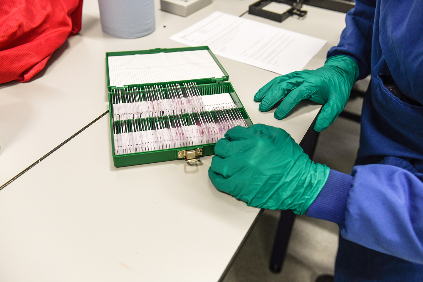
At the Institute, LCM is a highly used technique, supporting a wide range of research across our scientific programmes. In total, we have four Leica machines, each adapted to our specific research needs – supporting more than 40 users. Applications span cancer research, infectious disease and fundamental biology. The systems are continuously optimised to maintain high performance and reliability across this diverse user base.
Our LCM systems go beyond standard configurations, offering advanced capabilities that enable controlled environments for live or sensitive samples, seamless integration with imaging and annotation tools, and high-throughput workflows. These customisations make it possible to tackle complex experiments that standard instruments cannot accommodate.
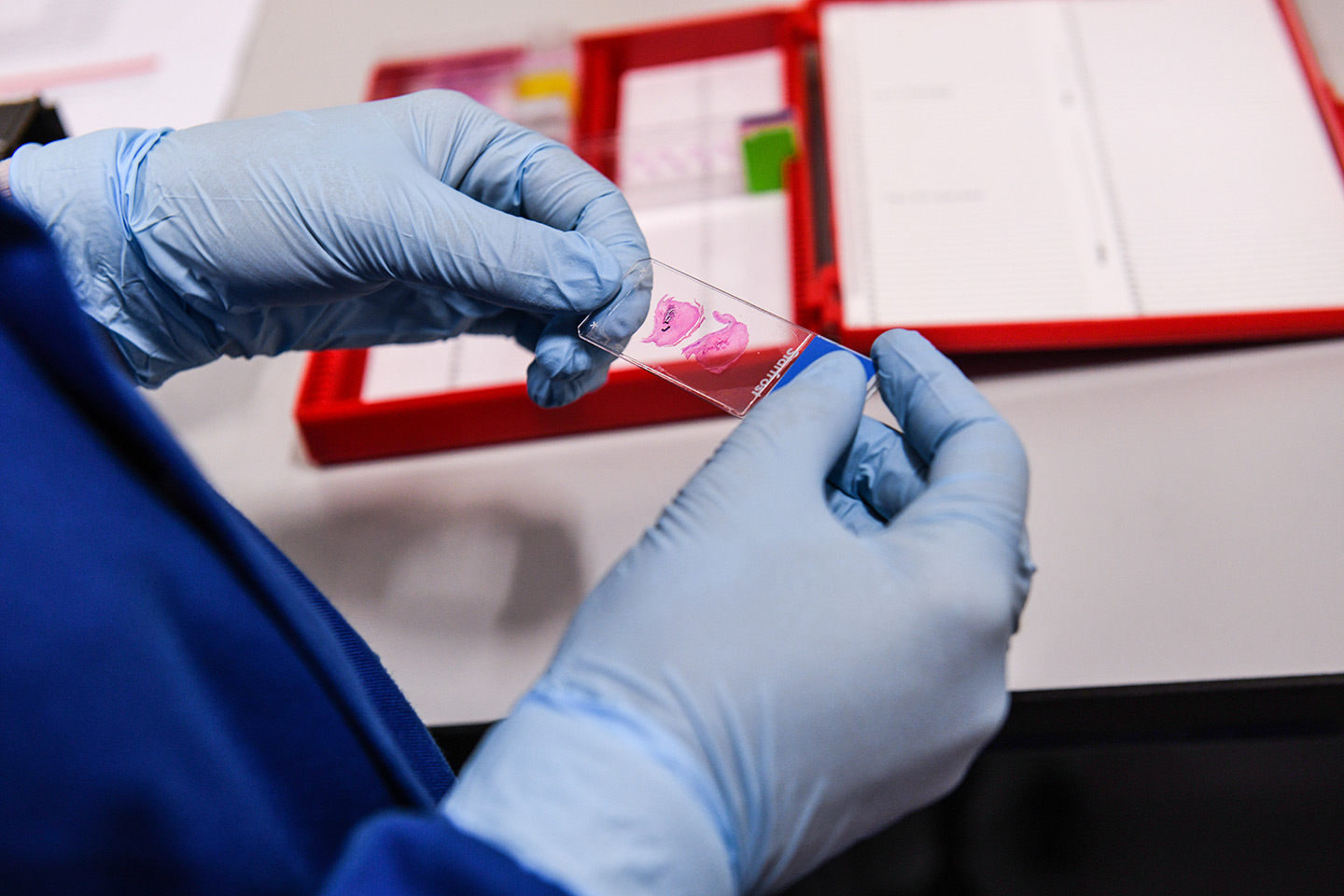
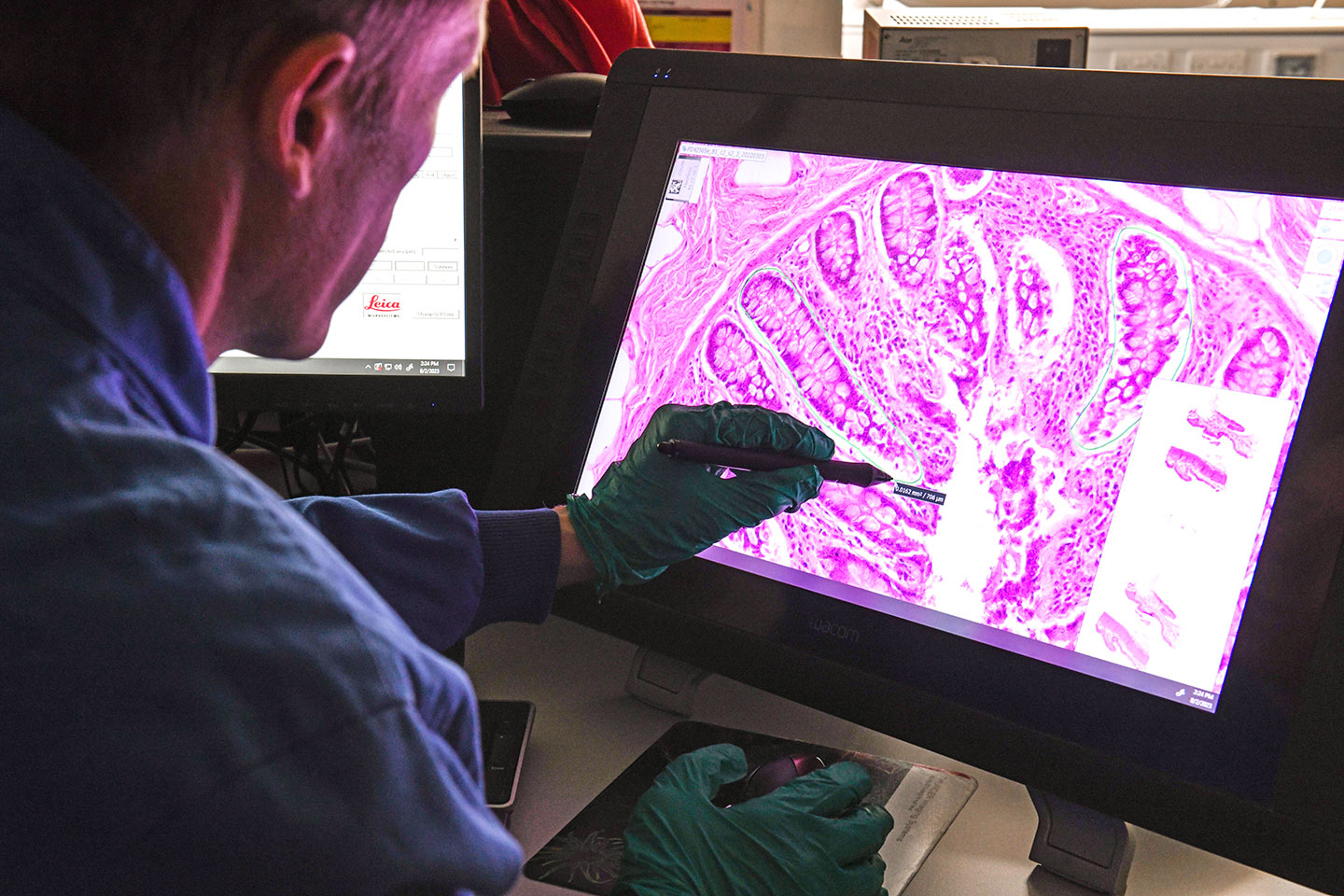
In practice, LCM is used to isolate specific cell populations from tissue while preserving their spatial context. For example, Professor Mike Stratton’s group is using LCM to isolate and study crypts – small gland-like structures – within the colon. His work is focussed on identifying the patterns of mutations that are driving the rise in colorectal cancer. Each crypt is quite unique because it comes from a small group of stem cells and acts like its own ‘clone’ – meaning many of the cells inside it, share the same DNA changes. By studying one crypt at a time instead of mixing lots of cells together, researchers can clearly see which mutations belong to each clone. This makes it much easier to track how mutations build up over time and identify the ones that may lead to colorectal cancer, as well as understand how these clones grow and spread in the earliest stages of the disease.
RELATED SANGER NEWS STORY
Sanger scientists receive major grant to study rise of early onset colorectal cancer
Professor Sir Mike Stratton and Dr Trevor Lawley, together with Dr Paul Brennan of the International Agency for Research on Cancer (IARC) have been awarded a significant grant, enabling their team to investigate the causes of early onset colorectal cancer.

LCM can also be used to study conditions outside of cancer. For example, Dr Iñigo Martincorena and his team recently used LCM to precisely isolate small clusters of immune cells, called lymphocytic infiltrates, from autoimmune thyroid samples. This careful isolation allowed them to apply a highly sensitive sequencing method called whole-exome NanoSeq to look for genetic changes, specifically driver mutations, that might contribute to autoimmune thyroid disease.
RELATED SANGER NEWS STORY
Improved genetic tool reveals hidden mutations that can drive cancer
Researchers have refined a powerful DNA sequencing tool that can uncover hidden mutations that occur naturally in our bodies as we age. In the largest study to date, they have used the tool to provide insights into the earliest steps of cancer development and the role of mutations in healthy tissue.

Thanks to this precise approach, the researchers could detect even very rare mutations in individual cells, providing strong evidence that somatic mutations can play a role in diseases beyond cancer. Using this method, the team was able to map out a surprisingly detailed picture of how these mutations accumulate and evolve in the thyroid during autoimmune disease, revealing a rich and dynamic landscape of somatic evolution in affected tissues. These findings will hopefully open new avenues for understanding disease development, identifying early warning signs and ultimately developing targeted therapies.
RELATED SANGER NEWS STORY
Hidden mutations in immune cells linked to autoimmune disease
New research suggests that autoimmune diseases may be driven by DNA mutations in immune cells that remove the natural brakes on the immune system. It reveals a previously hidden role for somatic mutations — DNA changes acquired throughout life — in diseases beyond cancer
The use of this technology extends beyond humans, including Inigo’s work with Dr Alex Cagan when he was a post-doc at Sanger. They previously used this technology to study small groups of intestinal crypts from different animals – including a giraffe and lemur – and at different ages, rather than focussing on single cells. This made it possible to uncover broader patterns of mutation and, as shown in their Nature paper, to compare how mutations accumulate across species – revealing that animals tend to reach a similar overall number of mutations by the end of life, even though they age at very different rates. This has led Dr Cagan to establish his own group at the University of Cambridge, and to them purchasing their own LCM system. The team is now expanding this work by using LCM on a wider range of human and mammalian tissues, as well as in other animals.
RELATED SANGER NEWS STORY
Mutations across the animal kingdom shew new light on ageing
Sanger researchers analysed genomes from 16 species of mammal, from mice to giraffes. The authors confirmed that the longer the lifespan of a species, the slower the rate at which mutations occur, lending support to the long-standing theory that somatic mutations play a role in ageing.
Where is LCM going?
LCM is a relatively mature technology, but its impact is still growing as it becomes combined with newer approaches in molecular and spatial biology. Rather than changing fundamentally, its power lies in how it works alongside and strengthens other techniques.
One of the most important developments is its integration with spatial biology, particularly spatial transcriptomics. These technologies can map gene activity across whole tissue sections and highlight regions or cell groups of interest. LCM can then be used to pick out those exact cells, enabling much deeper analysis. Together, this allows researchers to move from a broad ‘map’ of the tissue to very detailed, high-resolution molecular information.
RELATED SANGER BLOG
Ways Sanger is applying spatial transcriptomics to our science
Imagine being able to pinpoint exactly where in an aggressive brain tumour certain genes are turned on – like mapping a city’s most active neighbourhoods at rush hour. That is the promise of spatial transcriptomics, breakthrough technologies that are changing how scientists understand tissues, health and disease, and development.

Another emerging direction is the expansion of LCM sample processing into multi-omics – an approach that integrates data from multiple molecular layers. Researchers at Sanger have been working collaboratively across a number of groups to obtain multi-omic data (DNA, RNA and methylation) from individual LCM cuts. For example, recent work from Dr Carl Anderson’s group in the Human Genetics programme looking at ulcerative colitis – a chronic inflammatory bowel disease – has used microdissected colonic crypts to generate paired DNA and RNA sequencing data from the same structures. This enables the direct linking of somatic mutations with the effects on gene activity at single-crypt resolution.
At the same time, workflows are becoming more advanced thanks to better software and system integration. New tools make it easier to mark areas of interest, send them directly to the LCM system and carry out collections more smoothly. These improvements are speeding things up, reducing manual work and improving precision.
Despite this progress, full automation is unlikely in the near future. LCM is still affected by practical factors like tissue quality, membrane behaviour and focus, which require human judgement. As a result, the field is moving towards semi-automated workflows that combine software support with expert input, bringing together efficiency and reliability.
“LCM offers micrometre accuracy, elegantly isolating target cell populations and facilitating groundbreaking research, quite literally, at the cutting edge of science.”
Paul Scott,
Technical Specialist, Wellcome Sanger Institute
References
- Leica Microsystems. 20 Years of Leica Laser Microdissection. May 2021 [Last accessed: June 2026]
- Emmert-Buck MR, Bonner RF, Smith PD, Chuaqui RF, Zhuang Z, Goldstein SR, Weiss RA, Liotta LA. Laser capture microdissection. Science. 1996; 274: 998–1001.


